Corneal bioengineering
A clear cornea is essential for clear vision. The “window” of curved transparent tissue that sits in front of the iris and pupil is responsible for the majority of focussing of light entering the eye, enabling sight. If injury, infection, or other conditions result in scarring or cloudiness of the cornea, a transplant may be needed to restore vision.

Currently, corneal repair typically involves transplanting generously donated human tissue. While this method has a long-standing record of safety and effectiveness, the one-to-one donor-to-recipient ratio means that for every surgery performed, 70 patients remain untreated, waiting for this life-changing procedure. To address the global shortage of donor corneal tissue, we are collaborating with colleagues from the University of Sydney, University of Wollongong, University of Melbourne, the Centre for Eye Research Australia, and New South Wales Health – Organ and Tissue Donation Service to bioengineer corneal transplant products. This collaborative research initiative, known as BIENCO (Bio-Engineered Cornea), aims to significantly improve the donor-to-recipient ratio and pave the way for advancements in personalized medicine.
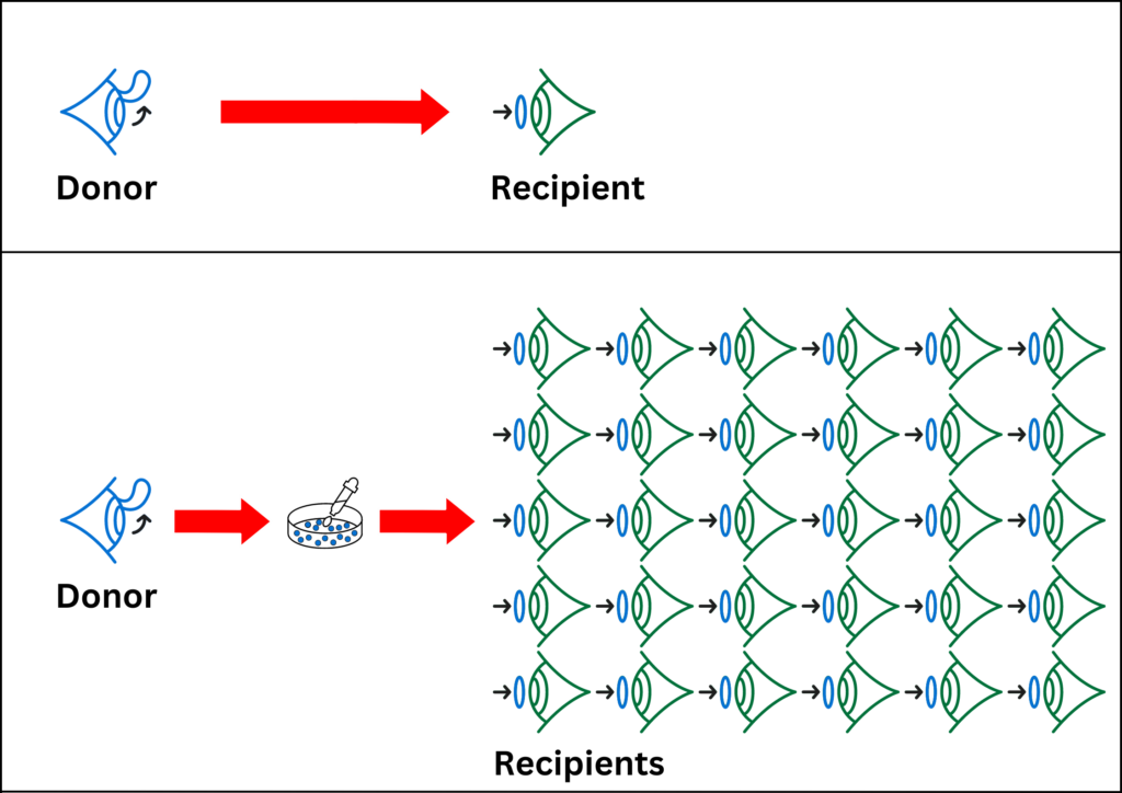
QUT’s contribution to the project is to apply our expertise in ocular cell culture to isolate and multiply the cells from donated eye tissues and test their compatibility with the proposed scaffolds for bioengineering the 3D corneal structure.
To stay up to date with all the latest news, you can follow the BIENCO consortium on social media, or check out the BIENCO Vision website.
Funding / Grants
- MRFF Medical Research Futures Fund (2023 - 2027)