In 1985, the first image of water diffusion in the living human brain came to life. Since then significant developments have been made and diffusion MRI has become a pillar of modern neuroimaging. Over the last decade, combining computational modelling and diffusion MRI has enabled researchers to link millimetre scale diffusion MRI measures with microscale tissue properties, to infer microstructure information, such as diffusion anisotropy in white matter, axon diameters, axon density, intra-cellular volume fraction, extra-cellular volume fraction. This microstructure information can provide significant insights on the diagnosis, progress and treatment of diseases, such as stroke, tumour, Alzheimer’s disease, and multiple sclerosis.
However, conventional modelling frameworks were developed assuming Gaussian diffusion of water molecules in the living tissue, which violates what is observed in the diffusion MRI signals. Hence, new modelling frameworks and computational methods that can capture non-Gaussian diffusion are needed urgently.
In this research domain, we focus on developing advanced diffusion MRI models to advance the field of microstructure imaging in the brain. Research in this domain is conducted with both domestic and international collaborators. Some of the projects within this research domain include:
- unifying several anomalous diffusion models under the continuous time random walk (CTRW) modelling framework;
- establishing mathematical links between the prevalent diffusional kurtosis imaging (DKI) model and our sub-diffusion model, which provides a new and more accurate way of estimating diffusional kurtosis;
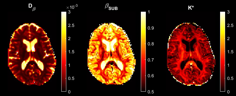
- evaluating how anomalous diffusion parameters change with age in the human brain corpus callosum;
- using parameters from time-fractional Bloch equation to improve human gray matter parcellation;
- evaluating if anomalous diffusion parameters can differentiate normal brain tissue and different layers in brain tumour.
Key Publications:
- Q. Yang, D.C. Reutens and V. Vegh. Generalisation of continuous time random walk to anomalous diffusion MRI models with an age-related evaluation of human corpus callosum. NeuroImage, 2022. (IF=6.556, ranked the top journal in the field of Neuroimaging).
- V. Vegh, S Moinian, Q. Yang, D.C. Reutens. Fractional order magnetic resonance fingerprinting in the human cerebral cortex. Mathematics 9 (13), 1549, 2021.
- Q. Yang, S. Puttick, Z.C. Bruce, B.W. Day, V. Vegh. Investigation of changes in anomalous diffusion parameters in a mouse model of brain tumour. Computational Diffusion MRI, 161-172, 2020.
Chief Investigators