Motivation: The increase in obesity prevalence is a major health concern in most developed countries. It has been suggested that increase in adiposity levels imposes a serious medical, social, economic, and psychological burden to society. In general, higher levels of body fat is strongly associated with higher rates of hypertension, cardiovascular disease, type 2 diabetes, depression and lower quality of life. Thus, obesity can directly cause a great impact on health care utilization and cost.
The type and location of fat storage within the body, rather than simply total body fat, might play a crucial role in the likelihood of developing cardiovascular and metabolic disease (Addison et al., 2014). In particular, there is a growing interests in intramuscular adipose tissue, which is the fat that accumulates beneath the musculoskeletal connective tissue, between muscle fibres or within muscle cells. It has been demonstrated that high levels of intramuscular fat are associated with an increase in metabolic risk factors (Therkelsen et al., 2013), reduced muscle function (which can lead to a cycle of increase in fat deposition due to a decrease in physical activity) and all-cause/cardiovascular mortality (Miljkovic et al., 2015). This is highly relevant to populations that typically exhibit high levels of intramuscular fat, such as stroke survivors, diabetic patients, spinal cord injured patients and the elderly. Thus, it is imperative that practical methods to quantify changes in intramuscular fat in clinical populations be developed.
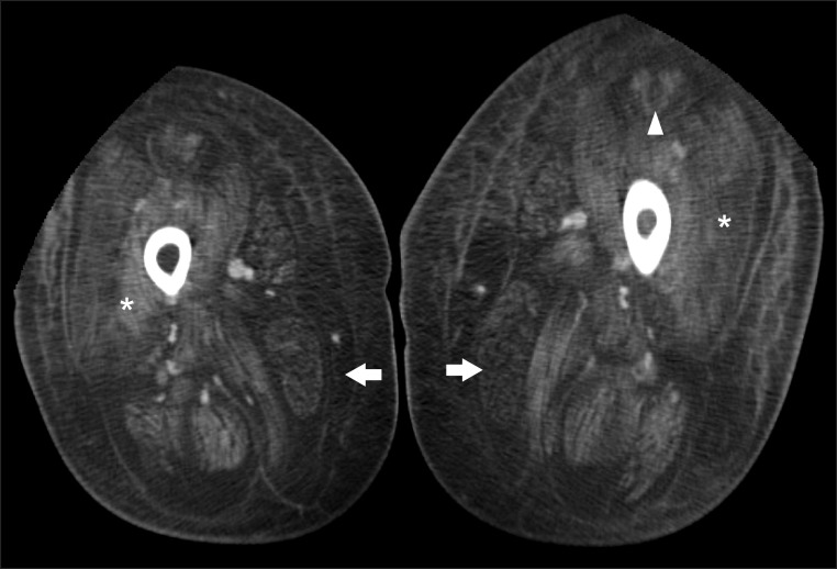
Outline: Intramuscular fat is typically measured using computed tomography (CT) or magnetic resonance imaging (MRI). Since these technologies are not always easily accessible, and rather costly, the assessment of intramuscular fat is not yet routinely performed in clinical practice and it is restricted to research practice. Alternatively, musculoskeletal ultrasonography is an imaging technique that is relatively low cost, does not deliver any unwanted extra dose to the patient and is portable. In particular, with ultrasound elastography, in addition to obtaining morphological information, we can quantify the absolute elasticity value of soft tissue structures and obtain useful quantitative information about the mechanical properties of tissues. Shear wave elastography is considered the most suitable type of ultrasound elastography for the musculoskeletal system. It is widely used for tendons, ligaments, and muscles. The purpose of the work proposed here is to investigate if shear wave elastography can be used to perform quantification/estimation (and possibly localization) of the amount of fat in the muscles.
Data and methods: Ultrasound and shear wave elastographic scans of relevant muscle districts in volunteers will be compared with MRI scans. Moreover, the same type of scans will be performed in fresh frozen cadavers at QUT Medical Engineering Research Facility (MERF) and on animal specimens (such as lamb legs for example). After dissection, comparisons will be performed between the acquired images and the actual presence, quantity and distribution of fat in the muscle. Scanning protocols and image analyses will be produced.
Skills required: The scope of this project can be adjusted to last approximately 12-18 months, and would be suited to a Honours or Masters student. Ideally the student will have a background in Medical Physics, Biomedical/Biomechanical Engineering/Exercise Science or similar.
The student will join an internationally recognized team of researchers in the area of ultrasound imaging and medical image processing. As such they will gain exposure to the clinical and research environments and build their experience in designing and developing complex research projects.
Expected output:
- A thesis
- 1-3 journal papers, depending on time-frame and scope
If you are interested in this topic and you are looking for further information, please contact me: email d3.fontanarosa@qut.edu.au, ph. 0403862724.
Check my START database entries: START
REFERENCES
Addison, O., Drummond, M. J., Lastayo, P. C., Dibble, L. E., Wende, A. R., McClain, D. A., & Marcus, R. L. (2014). Intramuscular fat and inflammation differ in older adults: The impact of frailty and inactivity. Journal of Nutrition, Health and Aging, 18(5), 532–538. https://doi.org/10.1007/s12603-014-0019-1
Miljkovic, I., Kuipers, A. L., Cauley, J. A., Prasad, T., Lee, C. G., Ensrud, K. E., … Zmuda, J. M. (2015). Greater Skeletal Muscle Fat Infiltration Is Associated With Higher All-Cause and Cardiovascular Mortality in Older Men. The Journals of Gerontology. Series A, Biological Sciences and Medical Sciences, 70(9), 1133–1140. https://doi.org/10.1093/gerona/glv027
Therkelsen, K. E., Pedley, A., Speliotes, E. K., Massaro, J. M., Murabito, J., Hoffmann, U., & Fox, C. S. (2013). Intramuscular fat and associations with metabolic risk factors in the framingham heart study. Arteriosclerosis, Thrombosis, and Vascular Biology, 33(4), 863–870. https://doi.org/10.1161/ATVBAHA.112.301009
Other Team Members